Dental Implant Diagram
About Dental Implant #280
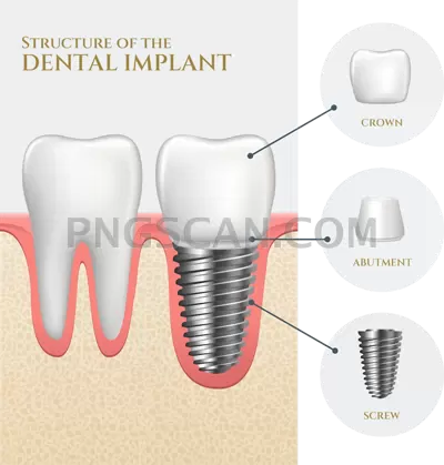
This dental implant diagram png shows what a patient never gets to see: the titanium post threaded into the jawbone, the abutment rising through the gum line, and the ceramic crown capping it off. The cutaway view slices through bone and tissue so each stage of the restoration reads at a glance, and the transparent background means nothing competes with it.
Dental clinics drop this into treatment pages and consent forms. Hygienists use it in chairside explainer slides when a patient asks why an implant costs more than a bridge. Dental supply catalogs, continuing-education decks, hygiene school worksheets, and orthodontic blog posts all pull the same asset because it explains the procedure faster than a paragraph of copy. You might also like Dental Implant.
At 400x419 pixels this sits comfortably as an inline figure or sidebar graphic rather than a hero image. If you need the labels, add them as live text beside the image instead of baking them into a raster at this size — text stays crisp, translates easily, and screen readers can actually reach it. Place it over a pale clinical blue or white and the natural bone tones stay legible. You might also like Dental Implant.
The file is a PNG with a true alpha channel, so the edges around the crown and bone section stay clean over any color or photo backdrop. Free license, no attribution required, cleared for both patient-education material and commercial clinic marketing. Explore more Dental Implant PNG images in our gallery.
Frequently Asked Questions
Answers about this Dental Implant #280 PNG — transparent background, size, download, and permitted use.
Three: the threaded titanium post anchored in the jawbone, the abutment connector above it, and the crown on top. Surrounding bone and gum tissue are shown in cutaway so you can see how the post integrates.
Yes, the free license covers print and clinical education use. At 400x419 pixels it prints well at roughly 1.3 inches wide at 300 DPI, so keep it as a small inline figure rather than a full-page illustration.
It will. Both keep the PNG alpha channel intact, so the implant sits directly on your slide or page color. Just avoid re-saving it as a JPG, which flattens transparency to white.
It is an illustrative diagram, not a scan or radiograph, so it works for explaining implant structure to patients and students. For clinical documentation or case records, use your own imaging.
More from the Dental Implant Collection


Related Transparent PNG Images

SUP Board

SUP Board 1500x1500

SUP Board natural

SUP Board 600x600

SUP Board 1000x1000